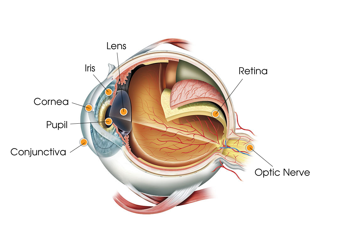
Conjunctiva: The thin, moist tissue that lines the eyelid’s inner surface, the conjunctiva helps protect the eye and prevents it from drying out.
Cornea: The eye’s front surface, the cornea covers the iris, pupil, and anterior chamber and is responsible for 70%of total focusing ability.
Iris: Located behind the cornea, is the colored part of the eye and contains the pupil. The iris adjusts the size of the pupil to control the amount of light entering the eye.
Lens: Located behind the pupil, the lens focuses light on the retina.
Optic Nerve: Transports visual signals from the retina in the back of the eye to your brain. It is made up of three-quarters of a million to more than a million nerve fibers.
Pupil: The black circle within the eye’s iris. The pupil controls how much light enters the eye by opening in darkness and closing as light increases.
Retina: Located at the back of the eye, receives images from the cornea and the lens and contains light-sensitive cells, called photoreceptors. There are two types of photoreceptors – rods and cones. Rods are more light sensitive while cones are more color sensitive.